Fayl:Respiratory system complete numbered.svg

Asl fayl (SVG fayl, asl oʻlchamlari 718 × 914 piksel, fayl hajmi: 507 KB)

Ushbu fayl Vikiomborga yuklangan boʻlib, boshqa loyihalarda ham qoʻllanilishi mumkin. Uning tavsif sahifasidan olingan maʼlumot quyida keltirilgan.

Taʼrif

[tahrirlash]

English: Note: See the version numbered to create or enhance one translation.

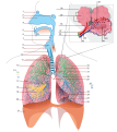
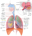
  • English: A diagram of the human respiratory system.
  • Català: Diagrama del sistema respiratori humà.
  • Español: Diagrama del sistema respiratorio humano.
  • Français : Schéma du système respiratoire humain.
Sanasi
Manba Oʻzimning ishim
Muallif LadyofHats, Jmarchn
Ruxsat
(Bu faylning takror foydalanilishi)
Public domain This work has been released into the public domain by its author, LadyofHats. This applies worldwide.

In some countries this may not be legally possible; if so:
LadyofHats grants anyone the right to use this work for any purpose, without any conditions, unless such conditions are required by law.

Other versions

[tahrirlash]

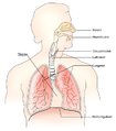
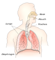
Simple
Complete
 
This W3C-unspecified vector image was created with Inkscape .

Translation

Language Text
en inglizcha

1: Paranasal sinuses (2: Frontal. 3: Sphenoid).

4: Upper respiratory tract: 5: Nose (6: Nasal cavity. 7: Nasal conchae. 8: Nasal vestibule). 9: Pharynx. 10: Larynx (11: Epiglottis. 12: Thyroid cartilage. 13: Cricoid cartilage). 14: Vocal folds.

15: Lower respiratory tract: 16: Trachea (17: Carina). Bronchi (18: Main bronchi. 19: Tracheal and bronchi rings. 20: Lobar bronchus (21: Superior. 22: Inferior. 23: Middle). 24: Lingular division bronchi). 25: Right lung (26: Superior lobe 27: Horizontal fissure. 28: Oblique fissure. 29: Middle lobe. 30: Inferior lobe). 31: Left lung (32: Superior lobe. 33: Apex of left lung. 34: Oblique fissure. 35: Cardiac notch. 36: Lingula of lung. 37: Inferior lobe).

38: Diaphragm.

39: Oral cavity. 40: Esophagus.

Respiratory lobule: 41: Connective tissue. 42: Alveolar sacs. 43: Alveolar duct. 44: Mucous gland. 45: Mucosal lining. 46: Pulmonary artery. 47: Pulmonary vein. 48: Capilllary beds. 49: Atrium. 50: Alveoli.

Annotations
InfoField
This image is annotated: View the annotations at Commons

Captions

Add a one-line explanation of what this file represents

Items portrayed in this file

tasvirlangan obyekt

dekabr 2007

Fayl tarixi

Faylning biror paytdagi holatini koʻrish uchun tegishli sana/vaqtga bosingiz.

Sana/VaqtMiniaturaOʻlchamlariFoydalanuvchiIzoh
joriy19:33, 2016-yil 14-fevral19:33, 2016-yil 14-fevral dagi versiya uchun tasvir718 × 914 (507 KB)JmarchnFixed error 43 arrow
00:35, 2016-yil 13-fevral00:35, 2016-yil 13-fevral dagi versiya uchun tasvir718 × 914 (507 KB)JmarchnRenumbered any bronchi
23:45, 2016-yil 12-fevral23:45, 2016-yil 12-fevral dagi versiya uchun tasvir718 × 914 (507 KB)JmarchnGrouping numbers
23:30, 2016-yil 11-fevral23:30, 2016-yil 11-fevral dagi versiya uchun tasvir718 × 914 (432 KB)JmarchnA lot of changes in upper respiratory tract and head
19:27, 2007-yil 13-dekabr19:27, 2007-yil 13-dekabr dagi versiya uchun tasvir800 × 900 (330 KB)LadyofHats{{Information |Description=numbered version of Image:Respiratory system complete.svg |Source=self-made |Date=dec 2007 |Author= LadyofHats |Permission=Public domain |other_versions=<gallery> Image:Respiratory system complete.svg|en

Bu faylga quyidagi sahifa bogʻlangan:

Faylning global foydalanilishi

Ushbu fayl quyidagi vikilarda ishlatilyapti:

Metama’lumot